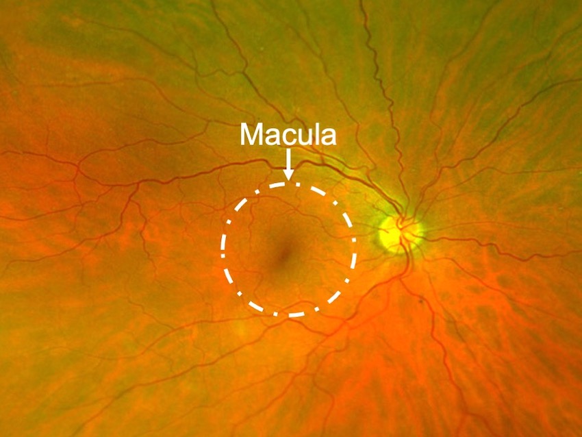
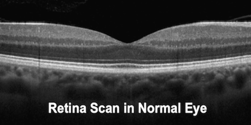
The retina is the thin layer of nerve tissue lining the back of the eye. The retina functions much like the film in a camera and transmits light signals to the brain. The central and most sensitive part of the retina is called the “macula”.
Vitreomacular Traction
What is vitreomacular traction (VMT)?
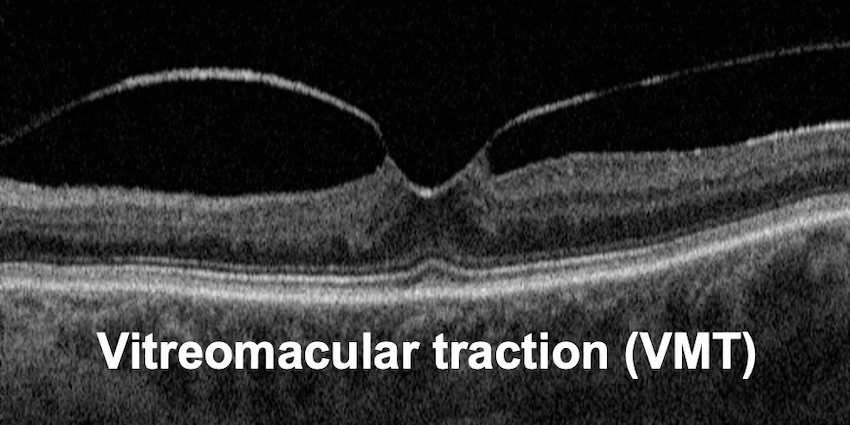
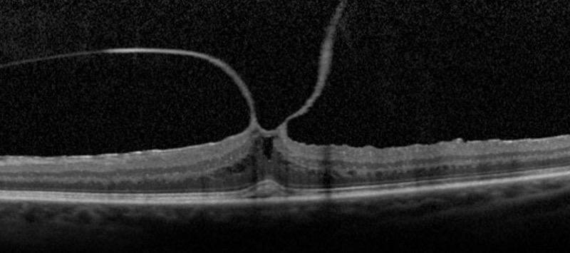
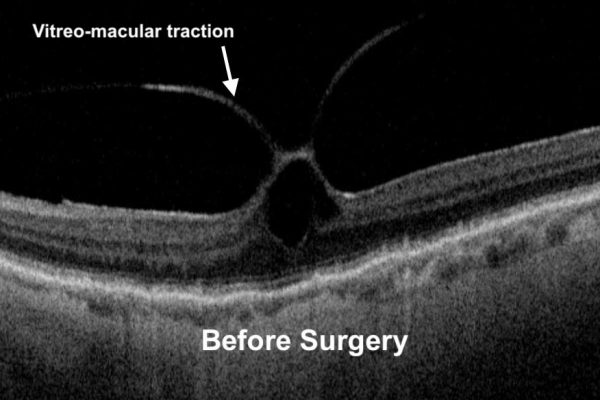
As we age, the vitreous (gel that fills the eye) shrinks, and sometimes it may pull away from the macula, leading to vitreomacular traction or VMT.
Vitreomacular traction often builds up slowly over time and may contract to cause wrinkling of the retinal surface, which may lead to blurring or distortion of your vision.
Symptoms
The majority of cases are mild and have a limited impact on vision. However, in some people, as the vitreous pulls more on the retina, this can damage the sensitive layers of the retina resulting in:
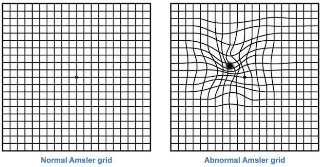
- Distortion of the image (straight lines appear wavy).
- Blurred vision particularly reading.
- Difference in the size of the image between both eyes.
Once diagnosed with vitreomacular traction it is important to regularly check your central vision with a certain grid called “Amsler grid”. This is a simple square containing a grid pattern and a dot in the middle. With vitreomacular traction, you may notice the lines are wavy or distorted.
How to diagnose?
If you have symptoms or distortion of blurry central vision, you should visit a retina specialist for an eye examination. This examination often involves:
- Visual acuity testing.
- Detailed back of the eye examination: often requires dilating eye drops to examine the central and peripheral retina.
- Retina photographs.
- Retina scans called Optical Coherence Tomography (OCT). This machine takes cross-section scans of the retina to identify the changes in the structure of the retinal layers.
What is the treatment?
In many patients, the vitreomacular traction is minimal and has only a limited impact on vision, therefore, no treatment is needed. However, in some people, the vitreomacular traction can be large causing significant visual disturbance making it difficult to carry out daily activities such as driving or reading. In such cases, the retina specialist may consider a treatment which is surgery.
The surgery is called “vitrectomy” which involves removing the gel inside the eye using very small instruments and release of the vitreomacular traction from the retina with the aid of fine micro-forceps.
Vitrectomy surgery is performed with the aid of local anaesthetic (small injection of anaesthetic solution around the eye) as day surgery. Your eye will be numbed and you will not feel any pain, although you may feel some pressure sensation during the surgery. The surgery often takes about 30-40 minutes. The surgery can be combined with cataract removal if you have a cataract (or if you are over 60 years).
Mr Ellabban will discuss with you the details of the surgery and advise you about the procedure.
Should I have surgery?
Mr Ellabban will discuss with you in detail about the surgery. The main reason to proceed with the surgery is to attempt to improve vision. If your symptoms are minimal and you are not aware of any visual problems, you might not need the surgery. However, if the distortion affects your ability to drive, read or perform important tasks, you should consider having the surgery.
If you have any queries about your suitability for vitreomacular traction surgery, you can Request a Call Back by filling out the form in the contact section.
Will my vision recover completely after the surgery?
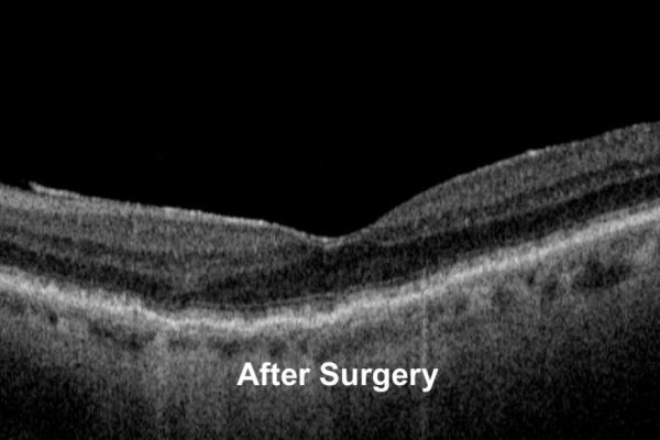
When the vitreomacular traction is released by surgery, you will usually notice an improvement in vision, particularly in the quality of vision. This usually takes a few weeks to reach the final improvement as the retina heals after the surgery. You will be followed with the aid of retinal OCT scans to monitor the healing process and restoration of the retinal layers. Also, the central distortion and blurring will gradually improve after surgery, however, it will not be completely restored as normal.
Following the surgery
You are advised to follow the instructions you were given after the surgery by Mr Ellabban and the nursing team. In general, there are important points to follow, after retinal surgery:
- Put the drops as you were instructed.
- Avoid rubbing your eyes for 2 weeks.
- Avoid heavy lifting and straining for 2 weeks.
- Avoid getting soap or shampoo or any chemicals into your eye for 4 weeks.
- Avoid swimming for 12 weeks.
- You must not drive until you reach the minimum legal standard of vision.
- It is normal for the eye to appear red or uncomfortable for a few days after the surgery which will gradually get better over time.
Mr. Ellabban will arrange a review after the surgery to monitor the healing process of the retina. You will be monitored with the aid of retinal OCT scans to assess the healing process and restoration of the retinal layers.
What are the risks of the surgery?
- Infection
- Prolonged Inflammation
- Bleeding
- Retinal detachment
- Increased eye pressure
- Distortion or alteration of vision
However, serious side effects are uncommon. There is a very small risk (less than one in 1,000) that you could lose sight in the eye as a consequence of the operation.
What if I don't have surgery?
As mentioned above, most cases of vitreomacular traction are mild and no treatment is required. The surgery is only indicated for selected cases where the vitreomacular traction has a significant impact on vision while performing daily activities like reading or driving.
If no surgery is performed, the vision will remain the same, however, in about 20-30 %, the vitreomacular traction may grow over time causing more distortion and blurring.
If you have any queries about your suitability for vitreomacular traction surgery, you can Request a Call Back by filling out the form in the contact section.
