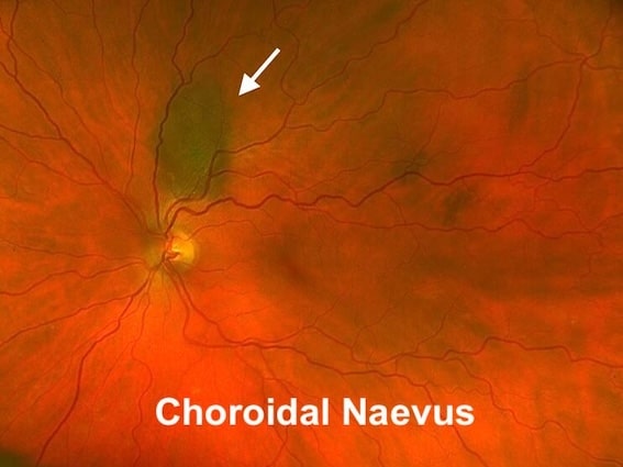
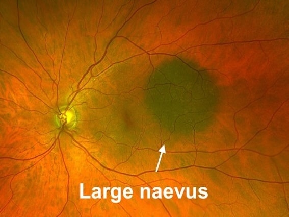
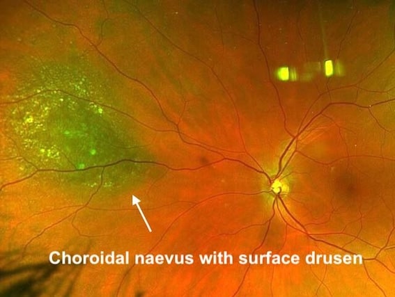
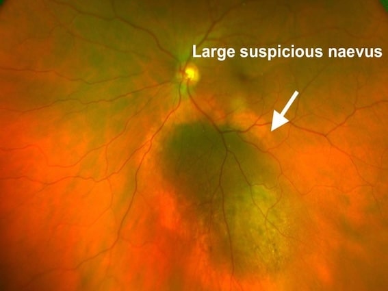
Choroidal Naevus (Freckle)

What is a Choroidal Naevus or Freckle?

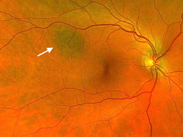
Choroidal naevus or freckle is a pigmented (darkly coloured) patch seen in the retina (back of the eye). It is similar to skin naevi (moles) that may be found in other parts of the body. The eyes contain cells that can produce pigment, similar to the skin,  and these cells can cause these “moles” to develop inside the eye.

Choroidal naevi are probably present at birth. They are often stable but may slightly grow in childhood and rarely beyond puberty.

Prevelance

Choroidal naevi are present in about 2-5 in 100 of the general population. They are often discovered by chance during an eye examination by the opticians or at the eye clinic.

Do I need treatment?

Choroidal naevi (freckles) are often left alone, as they are harmless and do not cause any problems. Occasionally, some naevi (freckles) may have suspicious features that will require monitoring, as a very small percentage of naevi can develop into a malignant (cancerous) lesion known as choroidal malignant melanoma.

Monitoring is done by taking pictures of the back of the eye and sometimes an ultrasound of the eye to monitor if there are any changes over time. It is often done once a year, but occasionally you may need to be reviewed more frequently at the first few appointments.

If you develop any of the suspicious features which increase the suspicion that melanoma could develop, Mr Ellabban may advise a referral to a regional centre for further assessment and to consider treatment. This is a very rare event. Otherwise, choroidal naevi are mostly harmless, although rarely they can affect vision in other ways for which treatment may be required.

Similar conditions

There are other conditions that may look similar to a choroidal naevus. The eye specialist often performs a detailed examination of the eye to rule out other similar conditions. Some of these conditions may affect your eyesight and need to be differentiated from a benign choroidal naevus.


These conditions are:

  • Choroidal melanoma.
  • Congenital Hypertrophy of the retinal pigment epithelium (CHRPE).
  • Subretinal or choroidal haemorrhage.
  • Retinal pigment epithelial tumours.
  • Choroidal hemangioma.
  • Choroidal granuloma.
  • Choroidal osteoma.
  • Posterior scleritis.
  • Sclerochoroidal calcification.
  • Solitary idiopathic choroiditis.